
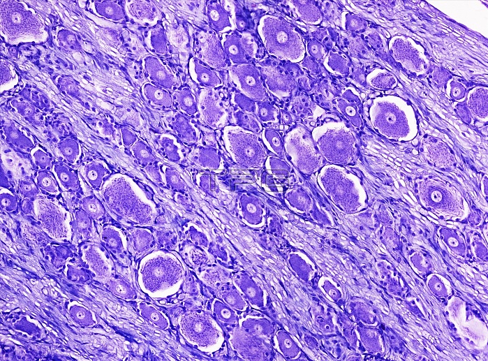
Light microscopy of a spinal sensory ganglion also known as a dorsal root ganglion. The ganglion is formed of a cluster of nerve cell bodies each with a central nucleus and densely stained cytoplasm (purple). Between the cell bodies are many myelinated axons that convey sensory signals from peripheral nerves to the spinal cord via the spinal ganglia. Magnification x100 when narrow width printed at 10 cm.
| px | px | dpi | = | cm | x | cm | = | MB |
Details
Creative#:
TOP15070391
Source:
達志影像
Authorization Type:
RM
Release Information:
須由TPG 完整授權
Model Release:
No
Property Release:
No
Right to Privacy:
No
Same folder images:
 Loading