
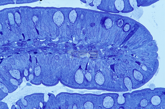
Small intestine lining. Light micrograph of a section through a villus from the lining of the human small intestine. A villus is formed of a layer of adjacent columnar epithelial cells (dark blue), which absorb nutrients from the gut lumen. The outer surface of the columnar cells is covered in tiny projections called microvilli. Interspersed with the columnar cells are goblet cells (pale blue), which secrete mucous onto the lining of the intestine to aid the movement of digested material through the gut. The interior of the villus is filled with blood vessels, connective tissue and smooth muscle cells. Magnification: x515 when printed 10cm wide.
| px | px | dpi | = | cm | x | cm | = | MB |
Details
Creative#:
TOP10220643
Source:
達志影像
Authorization Type:
RM
Release Information:
須由TPG 完整授權
Model Release:
N/A
Property Release:
N/A
Right to Privacy:
No
Same folder images:
 Loading