
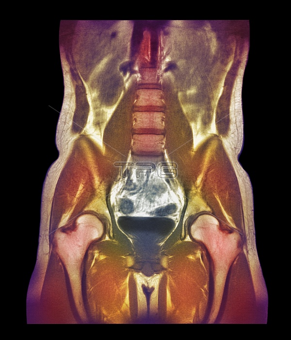
Abdominal scan. Coloured magnetic resonance imaging (MRI) scan of a woman's abdomen in coronal (frontal) section. The vertebrae (pale brown upper centre to centre) which form the spinal column are separated by the cartilaginous intervertebral discs (horizontal dark brown lines). Below the spine is the bladder (dark blue/black, lower centre), which receives urine from the kidneys (one seen at upper left, brown oval) and stores it before it is excreted. On either side of the bladder lie the femurs (thigh bones, pink/white, lower left and lower right, running to bottom). These are the longest and strongest bones in the body.
| px | px | dpi | = | cm | x | cm | = | MB |
Details
Creative#:
TOP10223680
Source:
達志影像
Authorization Type:
RM
Release Information:
須由TPG 完整授權
Model Release:
N/A
Property Release:
N/A
Right to Privacy:
No
Same folder images:
 Loading