
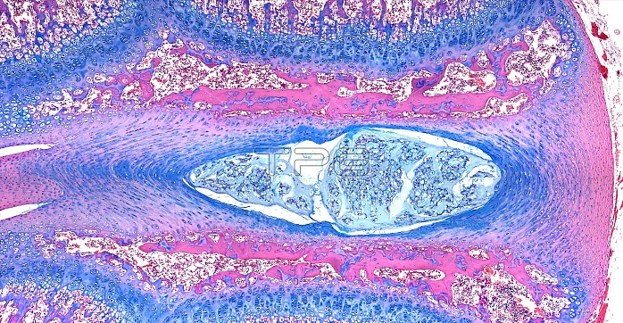
Light microscopy of an intervertebral disc. The disc is found between two vertebral bodies their bone tissue margins stained pink. The centre of the disc is formed of cells and a gel-like matrix, called the nucleus pulposus. It is a remnant of the head-to-tail axis of the early embryo (the notochord). Fibrocartilage rings (blue) form the outer margins of the disc, called the nucleus fibrosus. The disc acts like a cushion between the stacked vertebral bodies and resists compression but allows movements of each vertebra. A slipped disc' is an abnormal protrusion (herniation) of the central portion through a damaged region or tear of the fibrocartilage. Magnification x60 when narrow width printed at 10 cm.
| px | px | dpi | = | cm | x | cm | = | MB |
Details
Creative#:
TOP15070378
Source:
達志影像
Authorization Type:
RM
Release Information:
須由TPG 完整授權
Model Release:
No
Property Release:
No
Right to Privacy:
No
Same folder images:
 Loading