
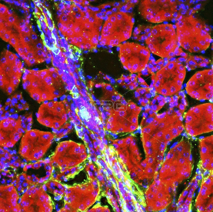
Kidney arteriole. Fluorescence deconvolution light micrograph of a section through an arteriole in kidney tissue, showing smooth muscle actin (green) and G-actin (red). Magnification: x400, when printed 10 cm wide.
| px | px | dpi | = | cm | x | cm | = | MB |
Details
Creative#:
TOP15298268
Source:
達志影像
Authorization Type:
RM
Release Information:
須由TPG 完整授權
Model Release:
No
Property Release:
No
Right to Privacy:
No
Same folder images:
actinanatomicalanatomyarteriolebiologicalbiologyblackbackgroundbloodvesselcellcellbiologycellscirculatorysystemcytologicalcytologyfluorescencedeconvolutionmicrographfluorescencelightmicrographfluorescentg-actinglobularhealthyhistologicalhistologyhumanbodykidneylightmicroscopemicroscopyno-onenobodynormalproteinproteinsrenalsectionsectionedsmoothmuscleactinvascularvessels
actinactinanatomicalanatomyarteriolebackgroundbiologicalbiologybiologyblackbloodbodycellcellcellscirculatorycytologicalcytologydeconvolutionfluorescencefluorescencefluorescentg-actinglobularhealthyhistologicalhistologyhumankidneylightlightmicrographmicrographmicroscopemicroscopymuscleno-onenobodynormalproteinproteinsrenalsectionsectionedsmoothsystemvascularvesselvessels
 Loading