
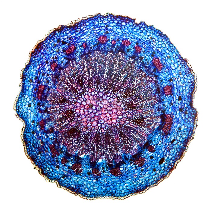
Fern (Osmunda regalis) petiole. Light micrograph of a section through a petiole (leaf stalk) from a royal fern, showing the epidermis (outer layer, red), chloroplasts (blue), endodermal cells (purple) and phloem sieve tubes (green). In the middle is the xylem, consisting of the metaxylem (large thick-celled, brown) and protoxylem (small thin-celled, brown). Magnification: x13 when printed 10 centimetres wide.
| px | px | dpi | = | cm | x | cm | = | MB |
Details
Creative#:
TOP16630737
Source:
達志影像
Authorization Type:
RM
Release Information:
須由TPG 完整授權
Model Release:
N/A
Property Release:
N/A
Right to Privacy:
No
Same folder images:
1anatomicalanatomybiologicalbiologybotanicalbotanycellcellschloroplastchloroplastscutoutcutoutscut-outcut-outscutoutcutoutsendodermalendodermisepidermalepidermisflorahistologicalhistologyleaflightmicrographlightmicroscopemetaxylemnatureno-onenobodyoneosmundaregalispetiolephloemplantplantsprotoxylemroyalfernsectionsectionedsievetubesinglestemtissuetubesvascularwhitebackgroundwildlifexylem
1anatomicalanatomybackgroundbiologicalbiologybotanicalbotanycellcellschloroplastchloroplastscutcutcut-outcut-outscutoutcutoutsendodermalendodermisepidermalepidermisfernflorahistologicalhistologyleaflightlightmetaxylemmicrographmicroscopenatureno-onenobodyoneosmundaoutoutspetiolephloemplantplantsprotoxylemregalisroyalsectionsectionedsievesinglestemtissuetubetubesvascularwhitewildlifexylem
 Loading