
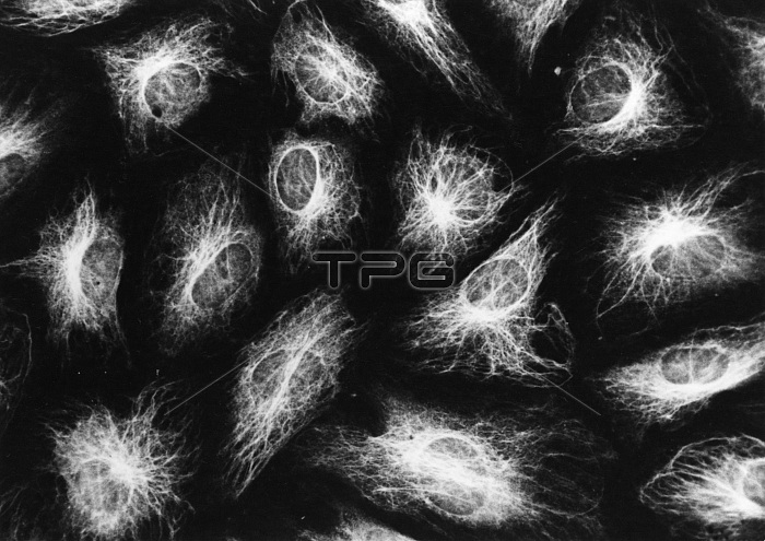
Photomicrograph of hamster N1L8 cells stained with antibody against a 58,000 dalton protein extracted from 10 nm filaments. Immunofluorescence makes it possible to get better information about the three-dimensional arrangement of cytoskeletal elements, by comparing the distribution of microtubules and 10 nm filaments in cells spread upon a tissue culture substrate.
| px | px | dpi | = | cm | x | cm | = | MB |
Details
Creative#:
TOP22220824
Source:
達志影像
Authorization Type:
RM
Release Information:
須由TPG 完整授權
Model Release:
N/A
Property Release:
No
Right to Privacy:
No
Same folder images:
 Loading