
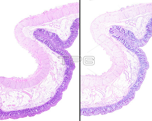
Composite image of two very low magnification light micrographs showing the large intestine (colon) wall stained with haematoxylin-eosin (left) and the PAS method (right). The mucosa layer show Lieberkuhn crypts with goblet cells stained with the PAS method.
| px | px | dpi | = | cm | x | cm | = | MB |
Details
Creative#:
TOP25663426
Source:
達志影像
Authorization Type:
RM
Release Information:
須由TPG 完整授權
Model Release:
N/A
Property Release:
N/A
Right to Privacy:
No
Same folder images:
 Loading