
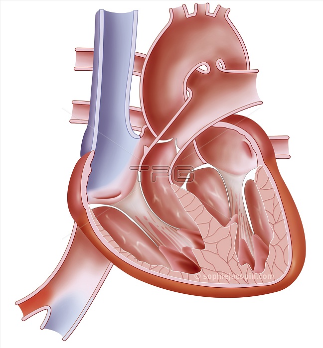
Heart of a fetus, pediatric anatomy, foramen ovale, arantius venous duct. Illustration representing the heart in a fetus, therefore before birth. Before birth, the right and left atrium communicate through the foramen ovale. This foramen closes at birth due to the pressure caused by the flow of blood. But the aortic arch also communicates with the pulmonary trunk at the level of the arterial duct. This duct closes 3 weeks after birth to become the arterial ligament. Another feature of the fetal heart is an additional blood vessel, the Arantius duct or duct. This vessel is used to transport venous blood from the placenta of the pregnant woman to the inferior vena cava of the fetus without having to pass through the liver. This helps transport oxygen from the umbilical vein much faster. The Arantius canal stops functioning a few minutes after birth. It will spontaneously close again during the first week of life and remain in the form of a residue: the venous ligament of the liver. Blue and red colors indicate oxygenation of the blood. Blue represents deoxygenated blood and red represents oxygenated blood.
| px | px | dpi | = | cm | x | cm | = | MB |
Details
Creative#:
TOP26089549
Source:
達志影像
Authorization Type:
RM
Release Information:
須由TPG 完整授權
Model Release:
N/A
Property Release:
N/A
Right to Privacy:
No
Same folder images:
 Loading