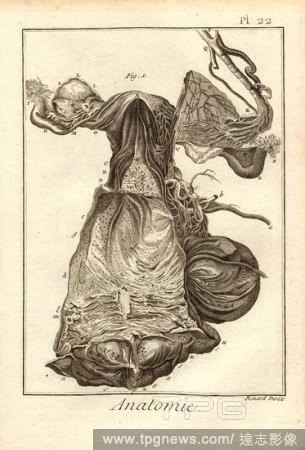
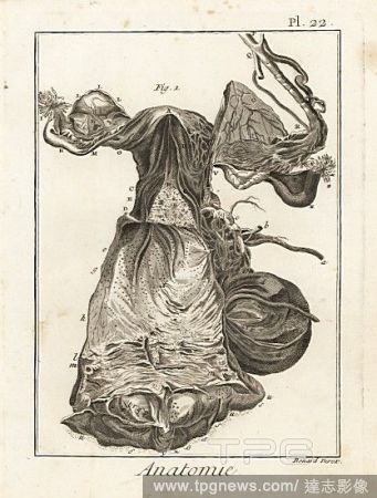
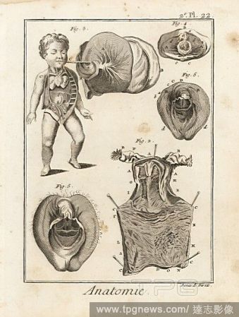
EditorialAnatomy, The matrix, according to Haller, Uterus, Signed: Benard direx, Pl. 22, to p. 48, Benard (dir.), 1778, Denis Diderot; M. d'Alembert: Encyclop?die, ou dictionnaire raisonn? des sciences, des arts et des m?tierss, des arts et des m?tiers. Gen?ve:...
EditorialLectures on the treatment of fibroid tumors of the uterus : medical, electrical and surgical : Martin, Franklin H. (Franklin Henry), 1857-1935.
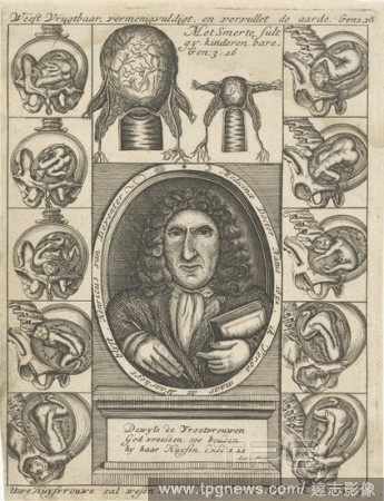
EditorialPortrait of Hendrik van Deventer and anatomical drawings of wombs with fetus, Portrait of Hendrik van Deventer in oval on base between anatomical drawings of wombs with fetus. Four Bible texts refer to the duty of the housewife to give birth to childre...
EditorialAnatomie, D?tails de la matrice, d'apr?s Haller & autres, Uterus, Signed: Benard direx, 2nd pl. 22, after p. 48, Benard (dir.), 1778, Denis Diderot; M. d'Alembert: Encyclop?die, ou dictionnaire raisonn? des sciences, des arts et des m?tierss, des arts ...
EditorialAnatomical drawing. Uterus and ovaries. Planches anatomiques du corps humain execute?es d’apre?s les dimensions naturelles. Paris France 1826. Anatomical print of the human body with natural dimensions. Uterus and ovaries. ANTOMMARCHI, C. France...
EditorialAnatomical drawing. Uterus and ovaries. . Planches anatomiques du corps humain execute?es d’apre?s les dimensions naturelles. Paris France 1826. Anatomical print of the human body with natural dimensions. Uterus and ovaries. ANTOMMARCHI, C. Fran...
EditorialAnatomical drawing. Male and female reproductive organs, Uterus, Ovaries, Penis and Testicles. Planches anatomiques du corps humain execute?es d’apre?s les dimensions naturelles. Paris France 1826. Anatomical print of the human body with natural di...
EditorialAnatomical drawing. Male and female reproductive organs - Uterus, Ovaries, Penis, and Testicles. Planches anatomiques du corps humain execute?es d’apre?s les dimensions naturelles. Paris France 1826. Anatomical print of the human body with natural ...
EditorialThe abdomen of a woman in the seventh month of pregnancy, showing section through uterus to reveal foetus. Copperplate engraving by Andrew Bell after an illustration by Jan van Rymsdyk from William Smellie's A Set of Anatomical Tables, Charles Elliot, ...
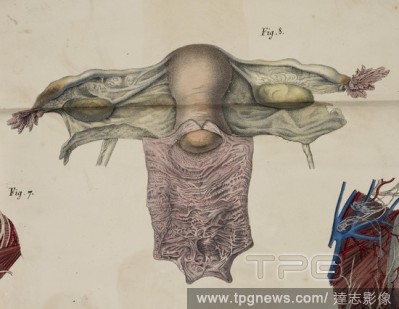
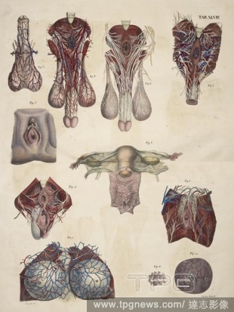
EditorialDetails of the uterus and female reproductive system including hymen, ovaries, Fallopian tubes, cervix, blood vessels, etc. Copperplate engraving by Robert Benard after an illustration by Albrecht von Haller from Denis Diderot's Encyclopedia, Pellet, G...
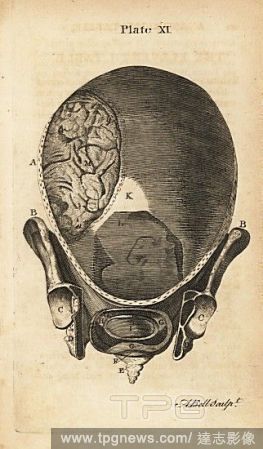
EditorialFront view of the gravid uterus at the start of labour. Copperplate engraving by Andrew Bell after an illustration by Jan van Rymsdyk from William Smellie's A Set of Anatomical Tables, Charles Elliot, Edinburgh, 1780.
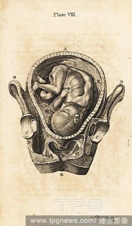
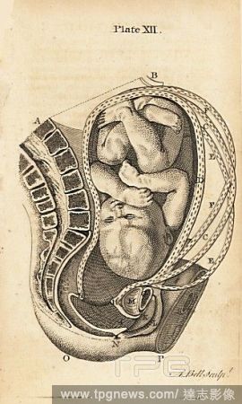
EditorialThe abdomen of a woman in the eighth or ninth month of pregnancy, showing section through uterus to reveal foetus. Copperplate engraving by Andrew Bell after an illustration by Jan van Rymsdyk from William Smellie's A Set of Anatomical Tables, Charles ...
EditorialDetails of the uterus and female genitalia, including vagina, hymen, clitoris, umbilical cord, foetus, ovaries, etc. Copperplate engraving by Robert Benard after an illustration by Albrecht von Haller from Denis Diderot's Encyclopedia, Pellet, Geneva, ...
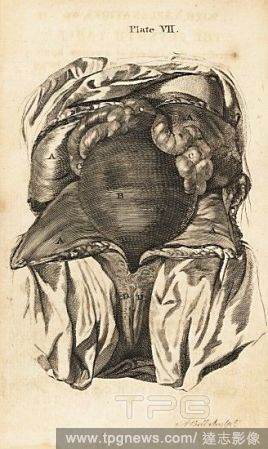
EditorialThe abdomen of a woman in the sixth or seventh month of pregnancy, showing uterus, intestines, labia pudendi. Copperplate engraving by Andrew Bell after an illustration by Jan van Rymsdyk from William Smellie's A Set of Anatomical Tables, Charles Ellio...
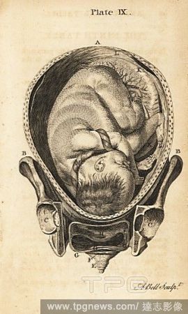
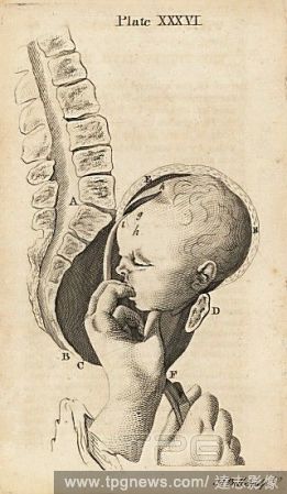
EditorialLateral view of the foetus in the uterus during advanced labour. Copperplate engraving by Andrew Bell after an illustration by Jan van Rymsdyk from William Smellie's A Set of Anatomical Tables, Charles Elliot, Edinburgh, 1780.
EditorialMethod of removing the severed head of a dead foetus from the uterus by hand and a curved crotchet. Copperplate engraving by Andrew Bell after an illustration by Jan van Rymsdyk from William Smellie's A Set of Anatomical Tables, Charles Elliot, Edinbur...
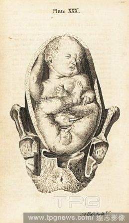
EditorialFront view of a foetus in breech birth position in the uterus. Copperplate engraving by Andrew Bell after an illustration by Jan van Rymsdyk from William Smellie's A Set of Anatomical Tables, Charles Elliot, Edinburgh, 1780.
EditorialView of the uterus in the second and third months of pregnancy. Copperplate engraving by Andrew Bell after an illustration by Jan van Rymsdyk from William Smellie's A Set of Anatomical Tables, Charles Elliot, Edinburgh, 1780.
EditorialFront view of the uterus in the vagina, and internal parts. Copperplate engraving by Andrew Bell after an illustration by Jan van Rymsdyk from William Smellie's A Set of Anatomical Tables, Charles Elliot, Edinburgh, 1780.


 Loading